This article is intended for healthcare professionals with training in ECMO management.
Recirculation is one of the most common and most underappreciated challenges in venovenous ECMO.
Understanding the mechanism is the prerequisite to recognising it at the bedside and managing it effectively.
What recirculation is
In VV-ECMO, blood is drained from the venous system, oxygenated through the membrane oxygenator, and returned to the venous system near the right atrium. The goal is for this oxygenated blood to enter the right heart, pass through the pulmonary circulation, and reach the systemic circulation.
Recirculation occurs when the return cannula tip is positioned too close to the drainage cannula tip. Freshly oxygenated blood, returned to the venous system, is immediately re-aspirated by the drainage cannula before it can enter the right heart. It cycles back through the ECMO circuit repeatedly, consuming the circuit's gas exchange capacity without contributing to the patient's oxygen delivery.
The clinical clue
The diagnostic signal is a striking discrepancy: high post-oxygenator saturation combined with persistently low patient arterial saturation. The circuit appears to be working perfectly; the patient continues to desaturate.
If the team interprets the high post-oxygenator value as evidence that the circuit is delivering adequate oxygen, the actual mechanism may not be recognised.
Management options
Repositioning the return cannula more proximally toward the tricuspid valve increases the distance between the return and drainage cannula tips, reducing the fraction of returned blood immediately re-aspirated.
Reducing circuit flow reduces the suction pressure at the drainage cannula, decreasing its tendency to draw back recently returned blood.
Optimising intravascular volume improves the filling of the venous system, increasing the volume of native venous blood available for drainage relative to recirculated flow.
Converting to a dual-lumen bicaval cannula addresses the positioning problem structurally when anatomy and clinical circumstances allow.
Relevance to Lifemotion circuit design
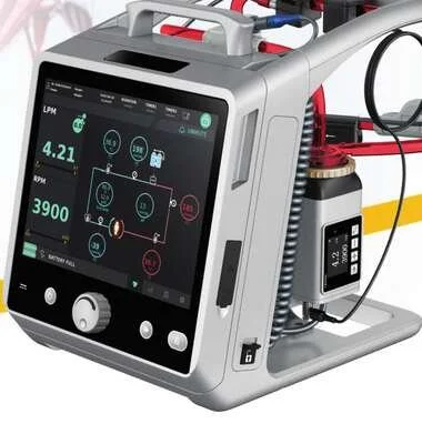
The Lifemotion ECMO platform's integrated monitoring including continuous venous SpO2, haematocrit, and haemoglobin on the console display provides the bedside information a team needs to identify the high-post-oxygenator / low-patient-saturation discrepancy characteristic of recirculation.
For ECMO programme teams managing VV-ECMO, understanding recirculation as a mechanism is part of the clinical competency base the programme should maintain.
Contact OHM Healthcare at sales@ohmhealthcare.com.au or visit lifemotionecmo.com.au for enquiries about the Lifemotion ECMO platform.
For healthcare professionals and healthcare organisations. Product availability and specifications subject to the locally approved Instructions for Use.
OHM Healthcare · sales@ohmhealthcare.com.au · 03 9115 7464 · lifemotionecmo.com.au

 Request Demonstration →
Service Support →