Cannulation — the placement of drainage and return tubes into the patient's vasculature — is the first and often most technically demanding step in initiating ECMO. The choice of cannulation strategy depends on the ECMO type (VV vs VA), clinical urgency, patient anatomy, operator expertise, and the intended duration and mobility of ECMO support.
VV ECMO cannulation approaches
Venovenous ECMO requires venous cannulation only. Common configurations:
- Femoro-jugular (standard two-cannula VV): drainage from the femoral vein (20–24 Fr); return via the right internal jugular vein (19–21 Fr). The most common configuration. Performed percutaneously under ultrasound guidance using Seldinger technique. Allows patient mobility when both sites are secured.
- Bicaval dual-lumen cannula (Avalon / Crescent): a single large-bore cannula (27–31 Fr) inserted via the right internal jugular vein with drainage ports in the SVC and IVC and return directed at the tricuspid valve. Requires echocardiographic positioning guidance. Eliminates the femoral cannula and allows greater patient mobility, including sitting and walking — important in awake ECMO and bridge-to-transplant patients.
- Femoro-femoral VV: both cannulae in the femoral veins; less commonly used due to higher recirculation risk
VA ECMO cannulation approaches
Venoarterial ECMO requires venous drainage and arterial return:
- Peripheral femoro-femoral (standard): femoral vein drainage (20–24 Fr); femoral artery return (15–17 Fr). Percutaneous; can be performed rapidly without cardiac surgery. Distal perfusion cannula (DPC) mandatory.
- Axillary artery cannulation: return via right axillary artery; allows antegrade (not retrograde) perfusion; avoids femoral ischaemia risk; permits patient ambulation; preferred in awake ECMO for ECPR or bridge-to-transplant
- Central cannulation: ascending aorta return + right atrial drainage; performed in the operating room; used post-cardiac surgery when chest is open, or when peripheral VA fails
- Transcaval approach: used in rare anatomical situations
Ultrasound-guided cannulation
All modern ECMO cannulation should be performed under real-time ultrasound guidance. This reduces procedural complications (vessel injury, haematoma, malposition) and confirms correct position before initiating full ECMO flow. Echocardiography (TOE or TTE) is used to confirm wire and cannula position in the heart and great vessels.
Surgical vs percutaneous approach
Percutaneous cannulation is now standard for peripheral ECMO at experienced centres, with surgical cut-down reserved for failed percutaneous attempts, severe obesity, or when direct vessel control is required. Central and axillary cannulation always require surgical access.
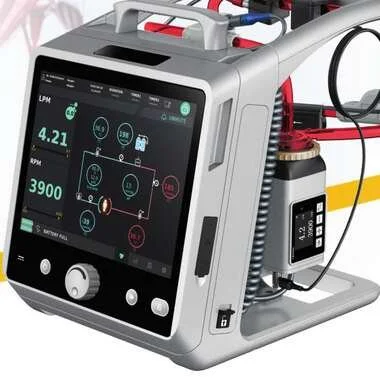
To learn more about the Lifemotion ECMO system — ARTG-listed and exclusively distributed across Australia and New Zealand by OHM Healthcare — visit us.
For healthcare professionals. Educational only — ECMO cannulation requires specific procedural training and credentialing.

 Request Demonstration →
Service Support →
Request Demonstration →
Service Support →