Neurological injury is among the most devastating complications of ECMO and a leading cause of death and disability in ECMO survivors. The brain is vulnerable in ECMO patients for multiple reasons: the underlying critical illness, haemodynamic instability before and during ECMO, anticoagulation-related bleeding, and embolic risk from the circuit. Systematic neurological monitoring is an essential component of ECMO care.
Types of neurological injury in ECMO
- Intracranial haemorrhage (ICH): the most common serious neurological complication; incidence 5–10% in adult ECMO; associated with anticoagulation; often fatal or leads to permanent disability
- Ischaemic stroke: from air or particulate emboli from the circuit, or from low cerebral perfusion pressure; less common than haemorrhagic stroke in adults
- Hypoxic-ischaemic encephalopathy (HIE): from pre-ECMO cardiac arrest or profound hypoxia; the degree of HIE before ECMO determines neurological prognosis in ECPR patients
- Critical illness polyneuropathy / myopathy: ICU-acquired weakness affecting peripheral nerves and muscles; detected on EMG/nerve conduction studies
- Delirium: very common in ICU ECMO patients; associated with prolonged sedation, sleep disruption, metabolic abnormalities
Risk factors for neurological injury
- Non-pulsatile ECMO flow (particularly VA ECMO) — impairs cerebral autoregulation
- Supratherapeutic anticoagulation
- Thrombocytopenia
- Haemolysis and gaseous microemboli
- Hypocapnia (over-aggressive sweep gas) — cerebral vasoconstriction
- VA ECMO with North-South syndrome — differential cerebral oxygenation
Neurological monitoring on ECMO
Surveillance strategies for neurological injury include:
- Daily neurological examination when sedation allows (GCS, pupillary response, gross motor function)
- Continuous EEG monitoring — to detect subclinical seizures, particularly post-cardiac arrest
- Cerebral near-infrared spectroscopy (NIRS) — non-invasive regional cerebral oxygenation monitoring; particularly useful in VA ECMO and North-South syndrome
- Brain CT — for any acute neurological change; MRI where feasible post-ECMO
Anticoagulation and ICH: a critical decision
When intracranial haemorrhage is diagnosed during ECMO, the team faces an immediate dilemma: stopping anticoagulation risks circuit thrombosis; continuing risks haemorrhage expansion. Decisions are made case-by-case, weighing the location and size of ICH, circuit age, indication for ECMO, and overall prognosis. Neurosurgical input is essential.
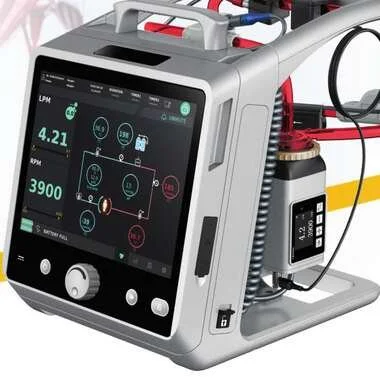
To learn more about the Lifemotion ECMO system — ARTG-listed and exclusively distributed across Australia and New Zealand by OHM Healthcare — visit us.
For healthcare professionals. Educational only — neurological complications of ECMO require multidisciplinary specialist input.

 Request Demonstration →
Service Support →
Request Demonstration →
Service Support →