Venoarterial ECMO bypasses the right heart and oxygenates blood returned to the arterial system — but the left ventricle (LV) remains in the circulation. When the LV is severely impaired and afterload is increased by ECMO return flow, the LV can dilate and distend (LV distension or "LV over-distension"), a complication that impairs recovery, increases the risk of pulmonary oedema and thrombus formation, and may ultimately require venting.
Why does LV distension occur on VA ECMO?
VA ECMO returns blood to the femoral artery in a retrograde direction, opposing the LV ejection. If the LV is very weak, it cannot generate enough pressure to eject against this increased afterload. Blood accumulates in the LV and left atrium (LA), pulmonary pressure rises, and pulmonary oedema may develop. On echocardiography, LV distension appears as a dilated LV with an aortic valve that fails to open (no LV ejection), and a distended LA.
Consequences of LV distension
- Pulmonary oedema and worsening respiratory function
- Reduced coronary perfusion (diastolic dysfunction)
- Impaired LV recovery — persistently elevated wall stress prevents myocardial rest
- Intracardiac thrombus formation (LV thrombus is a devastating complication)
Recognising LV distension
Daily echocardiography is essential during VA ECMO. Signs of LV distension include:
- LV end-diastolic dimension (LVEDD) increasing over time
- Absence of aortic valve opening
- Pulmonary artery pressure rising (if a PA catheter is in situ)
- New or worsening pulmonary oedema on chest X-ray
- Bright "smoke" in LV on echo (stasis)
LV venting strategies
The goal of LV venting is to decompress the LV and LA, reducing wall stress and pulmonary pressure. Options include:
- Intra-aortic balloon pump (IABP): reduces LV afterload and improves coronary perfusion; first-line unloading adjunct at many centres
- Impella micro-axial pump: directly unloads the LV by pumping blood from LV to aorta; highly effective but adds circuit complexity (ECMELLA configuration)
- Transatrial septostomy: balloon atrial septostomy creates an LA-to-RA communication, decompressing the left heart; used when other options are unavailable
- Surgical LV vent: placed intraoperatively through the LV apex or pulmonary vein
Australian practice
The combination of ECMO + Impella ("ECMELLA") is used at select Australian centres in refractory cardiogenic shock with LV distension. Careful haemodynamic monitoring and echocardiographic surveillance are central to managing this complex patient group.
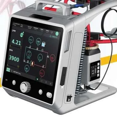
To learn more about the Lifemotion ECMO system — ARTG-listed and exclusively distributed across Australia and New Zealand by OHM Healthcare — visit us.
For healthcare professionals. Educational only — management of LV distension on VA ECMO requires specialist cardiac and ECMO expertise.

 Request Demonstration →
Service Support →
Request Demonstration →
Service Support →