Peripheral venoarterial ECMO requires a large-bore arterial return cannula — typically 15–17 French — in the femoral artery. This cannula can partially or completely occlude antegrade flow in the superficial femoral artery to the ipsilateral leg, causing acute limb ischaemia. Without timely recognition and intervention, limb ischaemia can result in irreversible muscle necrosis, fasciotomy, or amputation. It is one of the most preventable complications of VA ECMO.
Mechanism of limb ischaemia
The femoral artery divides into the profunda femoris and superficial femoral artery below the inguinal ligament. A 15–17 Fr arterial ECMO cannula occupies a significant proportion of the femoral artery lumen, reducing or eliminating antegrade flow in the leg. If collateral circulation is insufficient — as in patients with peripheral vascular disease or hypotension — the distal leg becomes ischaemic.
Prevention: the distal perfusion cannula (DPC)
The standard of care at high-volume ECMO centres is placement of a distal perfusion cannula (DPC): a smaller (5–7 Fr) sheath inserted antegrade into the superficial femoral artery, connected by side-port to the arterial return limb of the ECMO circuit. The DPC provides continuous perfusion to the distal leg throughout the ECMO run and is the single most effective preventive measure for limb ischaemia.
DPC placement: Should be performed at cannulation or within 6 hours of initiation, under ultrasound guidance. Flow through the DPC is typically 100–200 mL/min — enough to maintain limb perfusion without significantly reducing total ECMO flow.
Recognising limb ischaemia
Clinical assessment of the ipsilateral (cannulated) limb should occur at every nursing assessment — at minimum every 2–4 hours. Assess the 6 Ps:
- Pain — difficult to assess in sedated/paralysed patients
- Pallor — compare with contralateral limb
- Pulselessness — Doppler flow assessment of the dorsalis pedis and posterior tibial
- Paraesthesia — numbness in awake patients
- Paralysis — inability to move foot/toes
- Poikilothermia — cold, mottled limb
Near-infrared spectroscopy (NIRS) over the calf can provide continuous non-invasive monitoring of regional oxygen saturation and detect ischaemia before clinical signs develop.
Management of established limb ischaemia
- Ensure DPC is patent and flowing — check connections and flush
- Optimise anticoagulation — reduce thrombotic contribution
- If DPC not in place, insert urgently
- Surgical or interventional vascular assessment if ischaemia persists
- Compartment syndrome: measure compartment pressures, fasciotomy if indicated
- Consider alternative cannulation strategy (axillary or central) if femoral ischaemia cannot be resolved
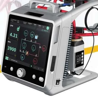
To learn more about the Lifemotion ECMO system — ARTG-listed and exclusively distributed across Australia and New Zealand by OHM Healthcare — visit us.
For healthcare professionals. Educational only — limb monitoring on VA ECMO must be embedded in nursing protocols at ECMO centres.

 Request Demonstration →
Service Support →
Request Demonstration →
Service Support →