VV-ECMO vs VA-ECMO: two configurations, two clinical pathways
ECMO is not a single therapy. It is a platform that can be configured in fundamentally different ways depending on what the patient needs. Veno-Venous and Veno-Arterial ECMO share the same core components — console, centrifugal pump, membrane oxygenator, circuit — but they operate in different haemodynamic territories and are used for different clinical indications. Understanding the distinction is essential for anyone involved in ECMO cannulation, management, or procurement.
How the circuit differs
In VV-ECMO, blood is drained from and returned to the venous circulation. The circuit sits in parallel with the right heart and lungs, providing gas exchange (oxygenation and CO₂ removal) without contributing to cardiac output. The patient's own heart must generate systemic blood flow — if cardiac function is impaired, VV-ECMO will not compensate for it.
In VA-ECMO, blood is drained from the venous side and returned to the arterial circulation, bypassing the heart and lungs entirely. This provides both gas exchange and haemodynamic support, making it suitable for patients whose cardiac output is insufficient to sustain life.
Veno-Venous ECMO
Cannulation
The two most common approaches:
- Bifemoral: drainage from the femoral vein, return via the right internal jugular vein (or contralateral femoral vein). The most established approach.
- Bicaval dual-lumen cannula (e.g. Avalon): a single cannula placed via the right internal jugular vein drains from the superior and inferior vena cavae and returns blood directed toward the tricuspid valve. Single-site cannulation; allows patient mobilisation.
Indications
- Severe acute respiratory distress syndrome (ARDS) — refractory hypoxaemia or hypercapnia despite optimised mechanical ventilation
- COVID-19 or influenza pneumonitis with refractory respiratory failure
- Bridge to lung transplantation
- Primary graft dysfunction following lung transplant
- Status asthmaticus (severe hypercapnic respiratory failure)
Key monitoring parameters
- Pre- and post-membrane gas analysis (assess oxygenator function)
- SvO₂ on the drainage limb (marker of oxygen delivery relative to demand)
- Recirculation fraction — blood returning from the circuit is immediately re-drained before entering native circulation, reducing effective gas exchange. Position and inter-cannula distance affect recirculation
Veno-Arterial ECMO
Cannulation
Two main approaches:
- Peripheral (femoral): drainage from the femoral vein; return via the femoral artery. The most common approach for emergency or percutaneous cannulation. Requires a distal perfusion cannula in the ipsilateral superficial femoral artery to prevent limb ischaemia.
- Central (surgical): drainage from the right atrium; return to the ascending aorta. Typically used in post-cardiotomy failure when the chest is already open.
Indications
- Refractory cardiogenic shock — post-myocardial infarction, post-cardiotomy, acute myocarditis
- Extracorporeal CPR (ECPR) — VA-ECMO deployed during refractory cardiac arrest
- Bridge to cardiac transplant or durable ventricular assist device (VAD)
- Refractory septic shock with severe cardiac dysfunction
- Massive pulmonary embolism with haemodynamic collapse
Key monitoring parameters
- Arterial pulse pressure — as native cardiac function recovers, the pulse pressure widens; a flat arterial waveform at adequate ECMO flow indicates severe native cardiac impairment
- Right radial SpO₂ or ABG — monitors for differential hypoxaemia (see below)
- Distal perfusion cannula flow — checks limb perfusion on the cannulated side
Differential hypoxaemia in peripheral VA-ECMO
One of the most clinically important hazards specific to peripheral VA-ECMO. Oxygenated blood is returned from the femoral artery and flows retrograde toward the aortic arch. If native cardiac function is partially preserved, the heart may eject deoxygenated blood (from impaired lungs) into the upper body and coronary arteries — while the ECMO circuit supplies the lower body with well-oxygenated blood.
This creates a haemodynamic divide at the mixing point in the descending aorta. The coronary arteries, cerebral circulation, and right arm receive poorly oxygenated blood; the lower body receives well-oxygenated ECMO output.
This phenomenon — sometimes called North-South syndrome or Harlequin syndrome — can result in myocardial and cerebral hypoxia despite apparently acceptable systemic parameters.
Detection: Continuous SpO₂ monitoring on the right hand (pre-ductal) and comparison with the SpO₂ probe used for standard monitoring. A significant right-arm SpO₂ drop relative to the lower body or pre-oxygenator circuit reading warrants investigation.
Correction strategies include:
- Optimising native lung function (ventilator adjustment)
- Increasing ECMO flow to push the mixing point further toward the aortic arch
- Adding a VV limb — converting to VAV-ECMO, where a return cannula in the jugular vein supplements oxygen delivery to the right heart and lungs
Selecting the right configuration
| VV-ECMO | VA-ECMO | |
|---|---|---|
| Gas exchange | Yes | Yes |
| Haemodynamic support | No | Yes |
| Cardiac output requirement | Native heart must function | Circuit provides cardiac output |
| Primary indication | Respiratory failure | Cardiac failure or arrest |
| Cannulation complexity | Lower (venous only) | Higher (arterial access) |
| Limb ischaemia risk | Minimal | Significant (distal perfusion cannula required) |
| Differential hypoxaemia | Not applicable | Yes — monitor right-arm SpO₂ |
Hybrid configurations
ECMO configurations are not always binary. Clinical situations sometimes require hybrid approaches:
- VAV-ECMO: a VV return limb added to a VA circuit — addresses differential hypoxaemia by supplementing pulmonary blood flow with oxygenated blood
- VVA-ECMO: additional drainage cannula in a VV circuit to augment cardiac support — used in mixed cardiorespiratory failure
- High-flow VV-ECMO with inotrope support: manages the respiratory component while medical therapy addresses the haemodynamic component
Clinical governance considerations for ANZ programmes
ECMO programmes in Australia and New Zealand that offer both VV and VA ECMO require defined cannulation protocols, a rostered team trained in each configuration, and governance pathways for configuration change mid-run. The ELSO General Guidelines provide a framework for programme structure; local adaptation to institutional volume and team composition is expected.
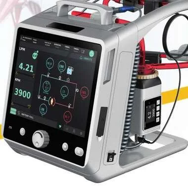
ARTG-listed ECMO systems must be suitable for both configurations. The Lifemotion ECMO system — distributed in Australia and New Zealand by OHM Healthcare — supports VV, VA, and hybrid ECMO configurations through a single integrated platform.
Reference: ELSO General Guidelines for all ECLS. Extracorporeal Life Support Organization. elso.org
To learn more about the Lifemotion ECMO system — ARTG-listed and exclusively distributed across Australia and New Zealand by OHM Healthcare — visit us.
For healthcare professionals. Educational only — ECMO configuration selection requires specialist clinical assessment and institutional governance frameworks.

 Request Demonstration →
Service Support →
Request Demonstration →
Service Support →