Weaning from venoarterial ECMO requires careful assessment of cardiac recovery and is more complex than weaning from VV ECMO. Unlike lung recovery — which can be tested by reducing flow and sweep gas — cardiac recovery must be demonstrated against the haemodynamic challenge of returning cardiac work to the native heart. This article reviews the principles and practical steps of VA ECMO weaning.
Assessing cardiac recovery before weaning
Daily echocardiography is the cornerstone of VA ECMO monitoring. Signs of cardiac recovery that suggest readiness for weaning:
- Aortic valve opening with each beat (return of LV ejection)
- Improvement in LV ejection fraction (LVEF) — typically toward ≥20–25%
- Reduction in LV end-diastolic diameter
- Return of pulsatility (widening pulse pressure)
- Falling lactate and improving mixed venous saturation (SvO₂)
- Reduction in vasopressor requirements
The formal weaning trial
Weaning is performed by stepwise reduction of ECMO blood flow while closely monitoring haemodynamics and echocardiographic parameters:
- Reduce ECMO flow from therapeutic levels (3–5 L/min) to 2.5 L/min — observe for 15–30 minutes
- Reduce to 1.5–2 L/min — observe MAP, HR, SvO₂, echo
- Reduce to 1–1.5 L/min (minimum safe flow to prevent circuit thrombosis without anticoagulation bolus)
- If haemodynamics remain acceptable (MAP ≥65, no significant deterioration in echo function), proceed to decannulation
Inotropic support (dobutamine, milrinone, levosimendan) is often titrated up during weaning to support the recovering heart. The ideal end point is haemodynamic stability on modest inotropic support at minimal ECMO flow.
Minimum flow caution: Flow below 1 L/min for more than a few minutes risks circuit clotting. If the trial cannot be completed quickly, an anticoagulation bolus is required, or the trial should be aborted and flow restored.
Decannulation
Once the weaning trial is successful, cannulae are removed. Femoral cannulae may be removed by vascular surgery (open or percutaneous with figure-8 suture technique). Direct pressure is held for 20–30 minutes; longer if anticoagulated. Limb vascularity is monitored closely post-decannulation.
When weaning fails
If the heart fails to recover, the clinical team must consider bridging strategies:
- Bridge to durable LVAD: transition from VA ECMO to a long-term ventricular assist device (LVAD) such as the HeartMate 3
- Bridge to transplant: in appropriate candidates, escalation to transplant listing
- Bridge to decision: time-limited continuation while awaiting neurological recovery or treatment of the underlying cause
- Goals-of-care discussion: if recovery or bridging is not possible, withdrawal of ECMO with palliative support
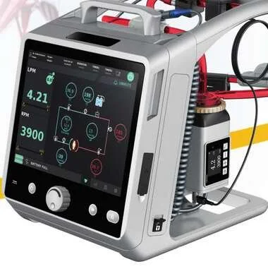
To learn more about the Lifemotion ECMO system — ARTG-listed and exclusively distributed across Australia and New Zealand by OHM Healthcare — visit us.
For healthcare professionals. Educational only — weaning decisions require multidisciplinary ECMO team input.

 Request Demonstration →
Service Support →
Request Demonstration →
Service Support →